

Tooth resorption
Resorption is a tooth slowly losing its own hard structure, often with no pain at all. A small-field 3D scan shows where it is and how far it has gone.
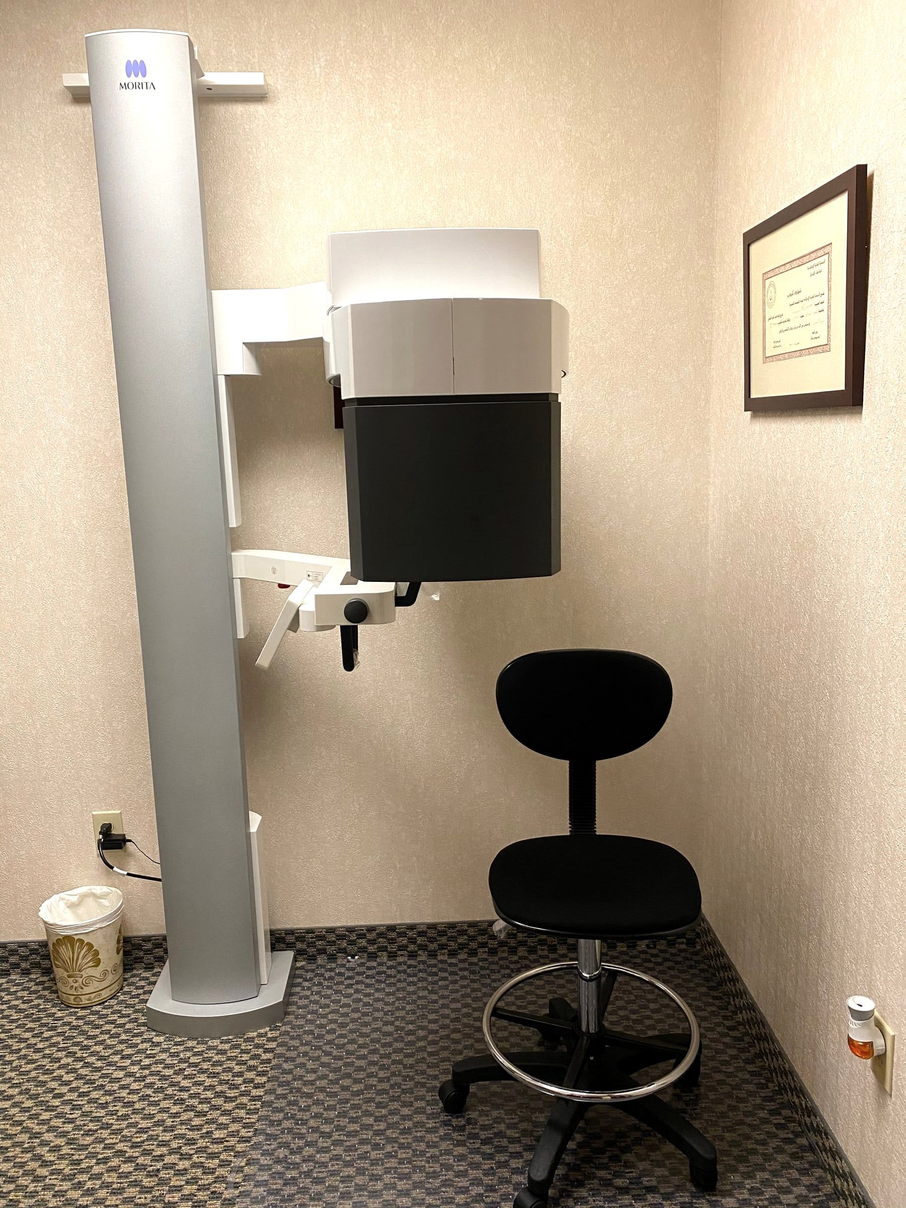
What resorption is
Resorption means the hard structure of a tooth, the dentin or the cementum that covers the root, is being broken down and lost. In children it is normal: baby teeth resorb as the adult teeth come in. In an adult tooth it is almost always a sign of a problem, and it is driven by inflammation next to the root or inside the tooth. When the cause of the inflammation is removed, inflammatory resorption stops.
Internal and external resorption are among the hardest cases in the specialty, and Dr. El-Refai treats both.
The kinds of resorption
Resorption is named for where it starts.
- Internal resorption starts inside the tooth, in the pulp, and widens the canal from within. It is uncommon and usually painless, and part of the pulp must still be alive for it to continue. Root canal treatment removes the tissue causing it, often with a medication placed inside the tooth first, and the outlook is very predictable and favorable.
- External inflammatory resorption starts on the outside of the root, next to inflammation caused by an infected or dead pulp, often after an injury. Cleaning and disinfecting the canals, usually with calcium hydroxide placed inside the tooth first, removes the cause so the resorption can stop.
- Replacement resorption, also called ankylosis, almost always follows an injury to the tooth. Bone slowly replaces the root as part of the body’s normal bone turnover. It cannot be stopped, but in adults a tooth with no symptoms can often be kept, sometimes for many years. In a growing child the tooth is watched closely, because it no longer moves with the growing jaw.
- External cervical resorption starts at the neck of the tooth, near the gum line, and can spread up or down the root and around it. It is usually painless and is often found by chance on an x-ray.
What may set it off
Cervical resorption has no single cause. The factors linked to it include orthodontic treatment, a past injury to the tooth, surgery near the neck of the tooth, internal bleaching of a root canal treated tooth, and grinding or clenching. In a 1999 study of 222 patients, orthodontic treatment was the only possible factor in 21.2 percent and an injury in 14.0 percent, while 15.0 percent had no identifiable factor at all. Studies like this show an association, not proof of cause.
In some teeth, a natural gap between the enamel and the cementum leaves a little dentin exposed at the neck of the tooth. The American Association of Endodontists puts this gap at 5 to 10 percent of teeth, and it may explain resorption in a tooth with no history of injury or surgery.
Resorption can also appear months or years after a tooth is injured, which is one reason an injured tooth needs follow-up visits.
Why a 3D scan matters
A flat x-ray can hide how large a resorption lesion is and where it sits. In a study of 115 teeth, regular x-rays underestimated the size of the lesions, and treatment plans changed significantly once a 3D scan was taken. The American Association of Endodontists and the American Academy of Oral and Maxillofacial Radiology recommend a small-field 3D scan as the preferred imaging to find resorption, tell internal from external resorption, and plan treatment.
Dr. El-Refai takes and reads every 3D scan herself and goes through it with you on the screen, so you can see where the resorption is and how close it comes to the nerve. More about our 3D imaging.
How it is treated
The treatment depends on the kind of resorption and how far it has spread.
- Root canal treatment for internal resorption, and for external inflammatory resorption caused by an infected pulp.
- Repair of a cervical lesion, from inside the tooth through a root canal, from the outside through the gum, or both.
- Monitoring with regular checks, when a lesion is stable or the tooth is best left alone for now.
- Removing the tooth, when the damage is too great to restore.
In a 2026 study of 296 teeth with cervical resorption, an estimated 85.7 percent were still in place after five years. Lesions that wrapped more than halfway around the root were linked to a higher chance of losing the tooth. We explain the options that fit your tooth and are honest about the odds. If a tooth is not a good bet, we will tell you.
Insurance and fees
In network with most dental PPO plans. We check your benefits and tell you what to expect before treatment. How payment works
Sources6 references
- American Association of Endodontists. Root resorption. Endodontics, Colleagues for Excellence, Spring 2025.
- Sousa Melo SL et al. AAE and AAOMR joint position statement, use of cone-beam computed tomography in endodontics, 2025 update. J Endod, 2026.
- Patel K, Mannocci F, Patel S. The assessment and management of external cervical resorption with periapical radiographs and cone-beam computed tomography, a clinical study. J Endod, 2016.
- Patel S et al. Clinical and radiographic features of external cervical resorption, an observational study. Int Endod J, 2023.
- Heithersay GS. Invasive cervical resorption, an analysis of potential predisposing factors. Quintessence Int, 1999.
- Patel S et al. The survival and success of teeth with external cervical resorption, a multi-centre, retrospective study. Int Endod J, 2026.

Relief starts with a call
Call or text, and we will find the soonest time that works. If you are in pain, tell us when you call, and we will do everything we can to see you the same day.