

In the room: the technology we use, and why
Endodontics is precise work in very small spaces. These are the tools that help us see more, treat more accurately and explain your tooth clearly, each chosen because published research supports it.
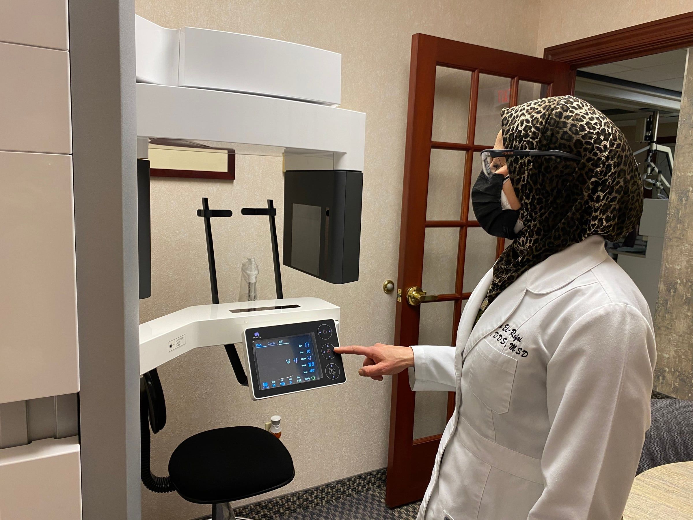
3D cone beam CT
Our Morita Veraview X800 is one of the most accurate, highest-resolution 3D scanners made for endodontics. It takes small-field 3D images at a fine 80-micron voxel size (maker's specifications). Limiting the scan to the area of interest keeps the dose low and the image sharp, because metal fillings and implants outside that area cannot streak the picture. It shows teeth, bone, sinuses and the structures around them in three dimensions: extra or missed canals, cracks and fractures, resorption, perforations, and the extent of infection that flat x-rays can hide. It is the foundation of every treatment plan, and it guides us if treatment becomes complicated. Dr. El-Refai takes and reads every scan herself, then goes through it with you slice by slice.
What the research shows
In a study of more than 1,500 teeth, a standard x-ray caught only about 55 percent of the infections a 3D scan found.
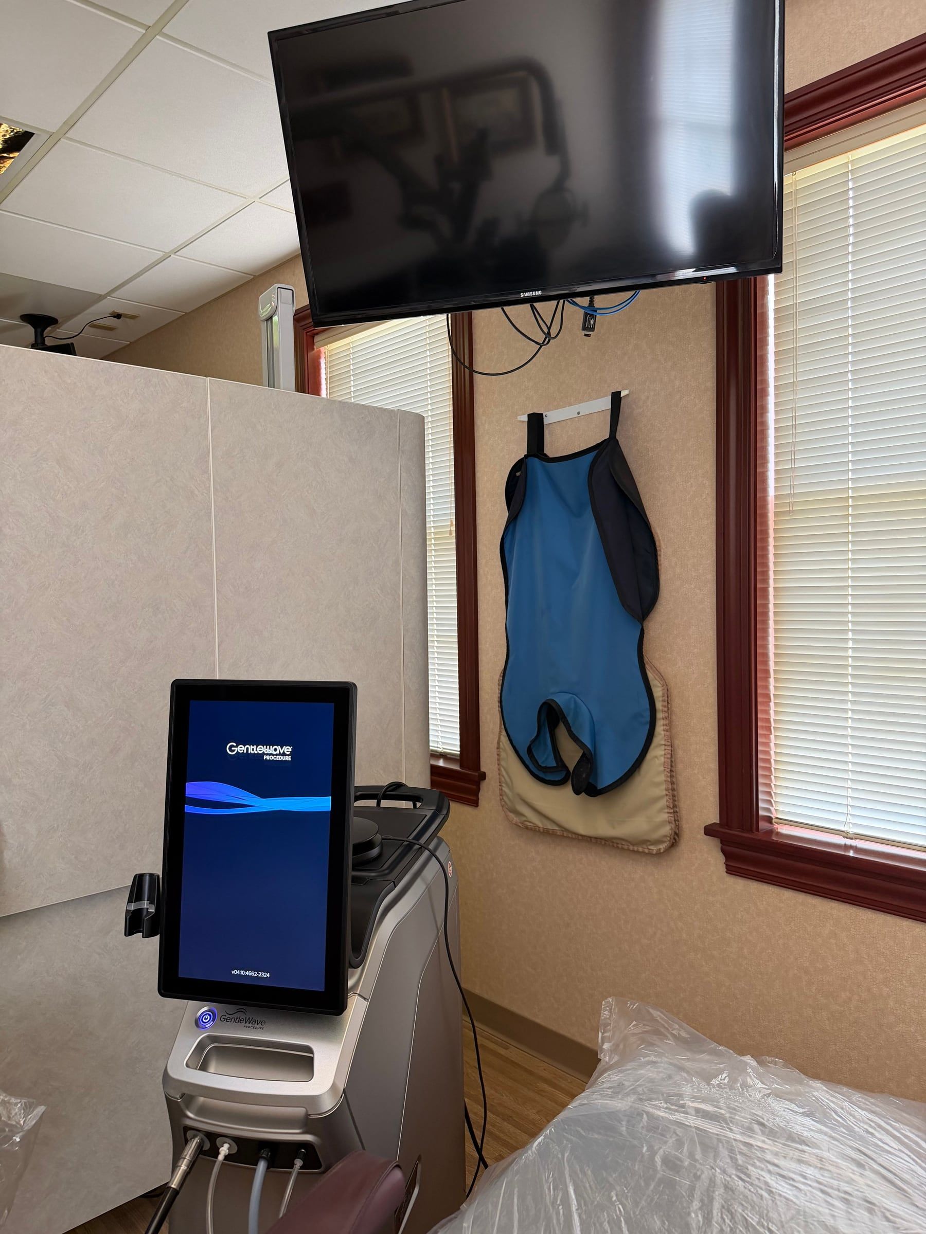
GentleWave
Fluid and broad-spectrum sound waves clean the whole root canal system, including small branches files can miss, and often allow treatment in a single visit. About GentleWave
What the research shows
In a multicenter clinical study, 97 percent of molars treated with GentleWave were healed or healing one year later.
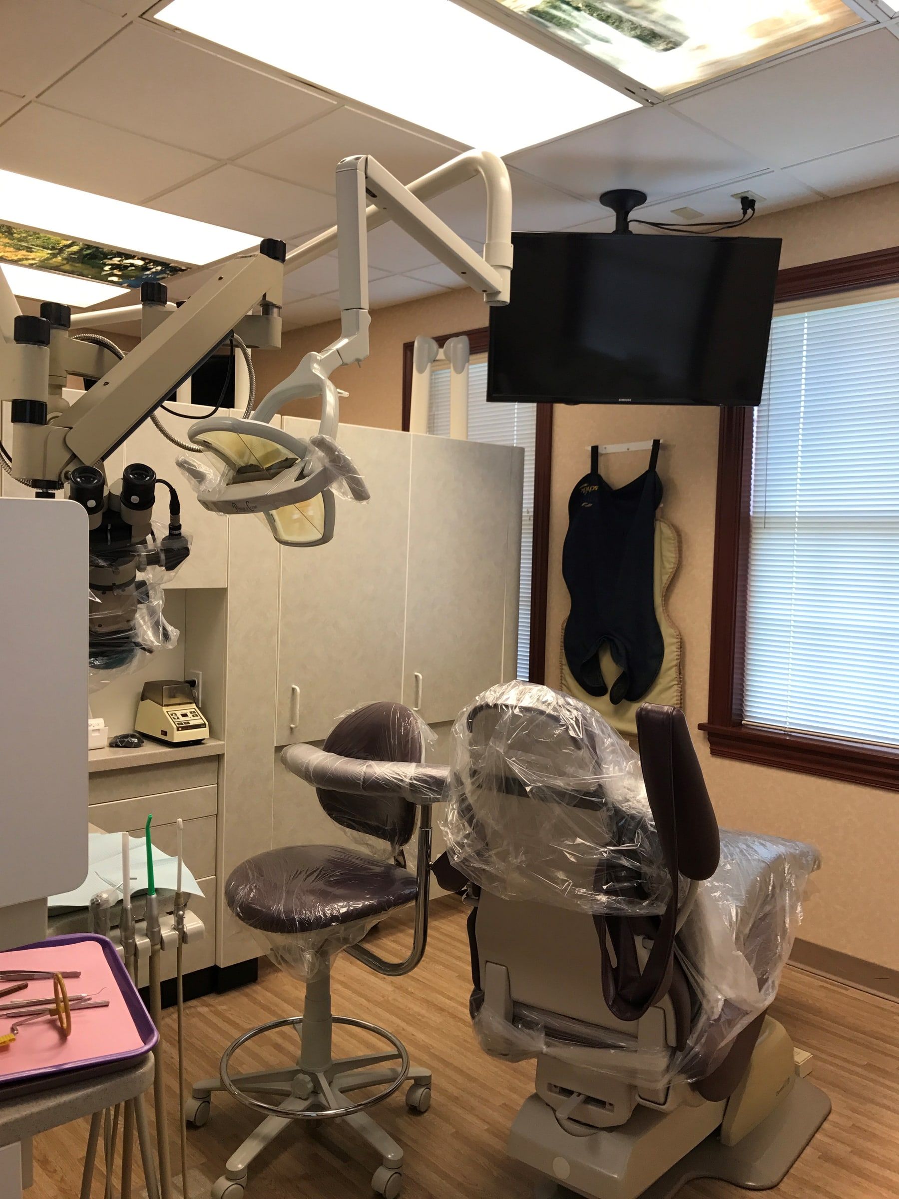
Surgical operating microscope
Every treatment room has one. High magnification and bright fiber-optic light make it possible to locate narrow, curved and calcified canals, spot fine cracks, and work with precision during retreatment and microsurgery. You cannot treat what you cannot see.
What the research shows
Endodontists using a microscope found the hidden second canal of upper molars in 57 percent of teeth, against 18 percent without magnification. In surgery at the root tip, modern microsurgery succeeded in 94 percent of cases, against 59 percent for older techniques.
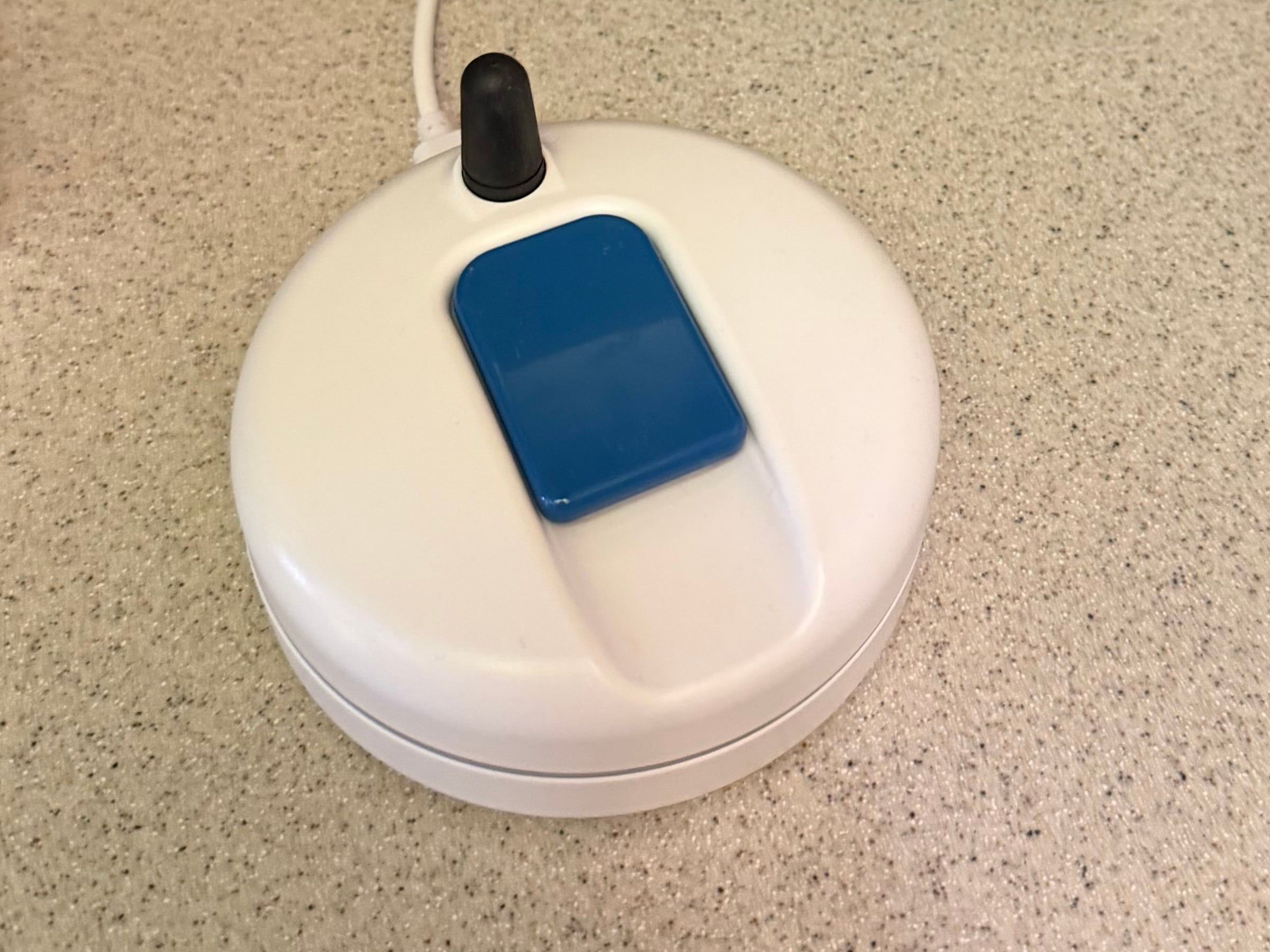
Wireless digital x-rays
Digital sensors need far less radiation than film, and images appear in seconds. Ours, the DC-Air, is the highest-resolution intraoral sensor by its maker's published measurements, keeping more than 70 percent at 5 line pairs per millimeter of the image's contrast (maker's specifications). That fine detail shows the narrow canals and small changes at the root tip that root canal care depends on. It is also wireless, so there is no cord in your mouth or to work around while the image is taken. We choose carefully which x-rays to take and when, following published guidelines, because they show what the eye cannot: decay between teeth, the level and health of the bone, the roots and the nerves inside them, cysts and other lesions, and damage after an injury. Images are stored digitally and can be sent to your dentist or your insurance company right away.
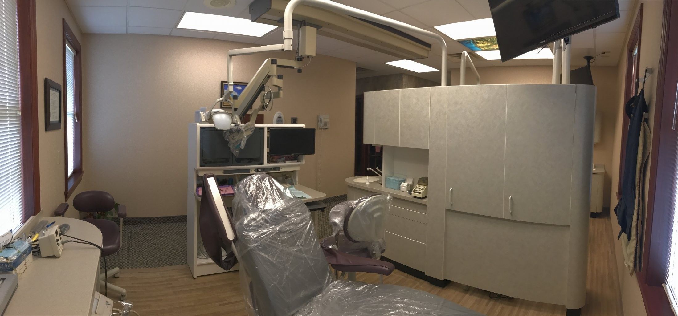
Instruments and materials
Ultrasonics. Every treatment room has an ultrasonic unit. Its fine diamond-coated tips vibrate up to 25,000 times a second, which lets us remove old posts and broken instruments, search for calcified canals, and prepare the end of the root for its filling during microsurgery.
Nickel-titanium instruments. The canals are cleaned and shaped with flexible hand and rotary instruments made of a nickel-titanium alloy, together with an antimicrobial irrigating solution that helps remove bacteria and tissue.
MTA. Mineral trioxide aggregate is a biocompatible ceramic material that sets in the presence of moisture and resists leakage, so it can be placed where the tooth is wet. We use it for pulp capping, repairing resorption and perforations, filling the root end during surgery, and forming a plug at the root tip in apexification.
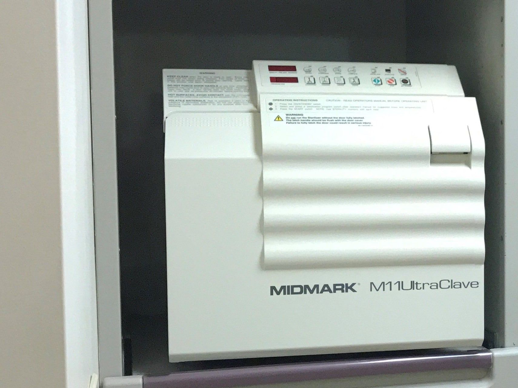
Sterilization
We follow the infection-control standards of the CDC, OSHA and the American Dental Association. Instruments are cleaned and autoclave-sterilized in a dedicated area after every use, with fresh barrier protection and personal protective equipment for every patient, hospital-grade disinfectants, single-use disposable materials wherever possible, and sterile saline in our water lines. Our sterilization equipment is monitored regularly.
Sources5 references
- Estrela C et al. Accuracy of cone beam computed tomography and panoramic and periapical radiography for detection of apical periodontitis. J Endod, 2008.
- AAE and AAOMR joint position statement, use of cone beam computed tomography in endodontics, 2015 update. J Endod, 2015.
- Buhrley LJ et al. Effect of magnification on locating the MB2 canal in maxillary molars. J Endod, 2002.
- Setzer FC et al. Outcome of endodontic surgery, a meta-analysis of the literature, part 1. J Endod, 2010.
- Sigurdsson A et al. 12-month healing rates after endodontic therapy using the novel GentleWave System. J Endod, 2016.

Relief starts with a call
Call or text, and we will find the soonest time that works. If you are in pain, tell us when you call, and we will do everything we can to see you the same day.