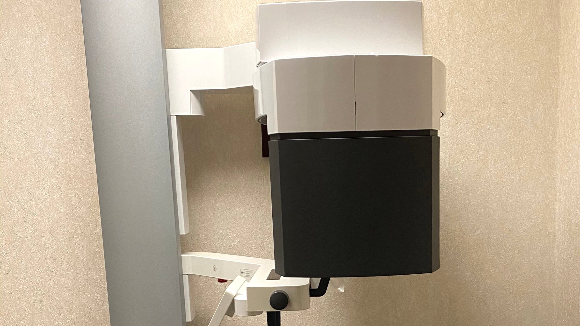
“But it doesn’t hurt.” We hear it often, and it is a reasonable thing to say when you are told you need a root canal. Pain is how most of us know something is wrong. With teeth, though, pain is not a reliable signal, and an infection can be present for a long time without any.
Infection without pain is common
Endodontists have a formal diagnosis for exactly this situation. In the American Association of Endodontists’ diagnostic terminology, asymptomatic apical periodontitis means inflammation and breakdown of the bone at the tip of the root, caused by a problem inside the tooth, that shows up on imaging but does not produce symptoms. A related diagnosis, a chronic apical abscess, often drains through a small pimple on the gum and may cause little or no discomfort.
This is why a problem tooth is often found on a routine x-ray at your dentist’s office rather than because it hurts.
Why a 3D scan sees more
A regular dental x-ray flattens a three-dimensional tooth into a single picture. Roots overlap, and the bone of the jaw can hide a lesion at the root tip.
A study of more than 1,500 teeth with endodontic infections compared regular x-rays with cone beam CT, the 3D scan endodontists use. The 3D scan found apical periodontitis significantly more often. The standard periapical x-ray caught about 55 percent of the lesions, and a panoramic x-ray only about 28 percent. Conventional x-rays reliably identified the problem mainly when it was already advanced.
Used when it adds something
More imaging is not automatically better. The joint position statement of the American Association of Endodontists and the American Academy of Oral and Maxillofacial Radiology calls for a small field of view and the lowest dose that answers the question, and for 3D imaging only when it adds information regular x-rays cannot. Common reasons include:
- a tooth with symptoms that do not match what a regular x-ray shows,
- complex root anatomy or a suspected extra canal,
- a previously treated tooth that is not healing,
- planning root-end surgery,
- some dental injuries and root resorption.
Why treat a tooth that feels fine
An infection that is not hurting today is still an infection, and the bone loss around the root can continue. Some stay quiet for years; others flare up without warning, sometimes as a painful swelling. Treating it on your schedule is usually easier than treating it in an emergency.
That said, not every spot on an x-ray needs treatment right away. A lesion that is already healing after earlier treatment may just need watching.
What you can expect here
Dr. El-Refai takes and reads your 3D scan herself, using a small-field cone beam CT, and goes through it with you on the screen. You see the roots, the bone and the infection, and we talk through your options, including when watching and waiting is reasonable. Learn more about our technology and root canal treatment.